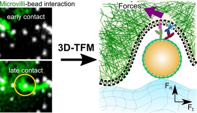
Functionalized Bead Assay to Measure Three-dimensional Traction Forces during T-cell Activation

Novel microscopy methods unravel the mechanical
interaction of T-cells with small particles.
Mechanical forces play a vital role for many biological mechanisms, such as the sensation of infected cells by T-cells. For example, T-cells (and other cells in our immune system) use mechanical forces to explore their environment, by pulling or pushing the other cells or tissue surfaces. These forces help T-cells to discriminate between pathogenic invaders and the body's own cells, during the so-called antigen recognition process. Recent studies show that the sensation of environment by T-cells is done via small "finger-like" protrusions of the cells, called microvilli.
In a recent study, an international Team of scientists with Prof. Enrico Klotzsch from Humboldt-Universität zu Berlin visualized the microvilli in action, where they exert local pushes and pulls at the surface of the antigen presenting surface.
Together with labs at EMBL Heidelberg, ETH Zurich, Medical Universities of Vienna, Charite and Vienna University of Technology, Prof. Enrico Klotzsch's lab has developed a platform, which allows to quantify the magnitude and direction of traction forces exerted locally by the T cell fingers and at the same time measuring the overall T cell activation status. This platform will find broad applications and contribute towards figuring out better treatments and improving patient health in adoptive immunotherapies.
Publication
Functionalized bead assay to measure 3-dimensional traction forces during T-cell activation, Nano Letters doi: 10.1021/acs.nanolett.0c03964
Contact
Prof. Dr. Enrico Klotzsch
Experimental Biophysics/Mechanobiology
Departmemt of Biology, Humboldt-Universität zu Berlin
